

Oh Young Bang, Jin Soo Lee, et al.
[Phase 1/2 Stroke Trial] Safety and Functional Recovery Following Intravenous Administration of Autologous Bone Marrow-Derived Mesenchymal Stem Cells in Patients with Ischemic Stroke
Research Summary
Five of 30 patients who had suffered a cerebral infarction in the middle cerebral artery territory and were left with severe disabilities received intravenous injections of mesenchymal stem cells cultured from their own bone marrow. The Badel Index, which measures the ability to perform activities of daily living, was higher in the treatment group than in the control group at both the 3-month and 6-month time points, and there were no serious adverse reactions related to the cells. However, the difference was not significant at the 12-month time point, and since only 5 patients received the treatment, it is too early to determine the efficacy. |
Study Results
Autologous bone marrow-derived mesenchymal stem cells were administered intravenously in two doses to patients with severe residual disability due to middle cerebral artery territory cerebral infarction. There was no specific primary endpoint; instead, the Badel Index (BI), which assesses activities of daily living, and the Modified Rankin Scale (mRS), which measures the degree of disability, were measured.
The Badel Index in the treatment group was higher than that in the control group at 3 and 6 months (P = 0.011, P = 0.017), but there was no significant difference at 12 months (P = 0.115). There was no difference in the Modified Rankin Scale at any of the three time points (P = 0.076, 0.171, 0.286). The range of change in the Neurological Deficit Score (NIHSS) was smaller than that of the Badel Index, and there was no difference in infarct volume (P = 0.661).
No immediate or delayed toxicity related to cell administration was observed. Over the course of one year, there were no deaths, recurrent strokes, or serious cell-related adverse events in the treatment group. Six months after administration, one patient developed cellulitis caused by athlete’s foot and was treated with antifungal medication. No tumors were detected on brain MRI.
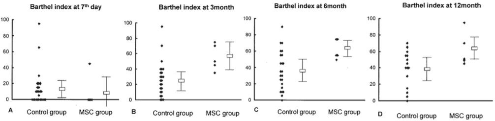 |
Figure. Comparison of the Badel index between the treatment group and the control group before (Day 7) and after (3, 6, 12 months) cell therapy. Original Figure 3, Ann Neurol.
Study Design
This was a randomized, controlled Phase 1/2 clinical trial conducted at a tertiary general hospital in Korea. Thirty patients aged 30–75 who had a confirmed lesion in the middle cerebral artery territory within 7 days of symptom onset and retained a disability score of 7 or higher on the NIHSS by the 7th day of hospitalization participated in the study. Five of the 10 patients assigned to the cell therapy group withdrew, so the final analysis included 5 patients in the treatment group and 25 in the control group. The control group received standard care only; no sham procedures were performed.
Assignment was performed on the 7th day of hospitalization by an independent investigator using a randomization table; however, subsequent trial procedures were not blinded. Evaluations were conducted seven times from the time of admission through week 52, and brain MRI scans were performed at week 52 on five patients in each group.
Cells Used and Administration Method
Autologous bone marrow-derived mesenchymal stem cells obtained from the patients’ own bone marrow were used. On the seventh day of hospitalization, 5 mL of bone marrow was collected from the posterior region of the ilium and cultured at Pamisell’s GMP facility; it took an average of 30.8 days (±5.5) to expand the cells to 1×10⁸ per patient. Pre-administration testing confirmed that over 91% of the cells expressed the CD105 surface antigen; the survival rate exceeded 95%, and there was no microbial contamination.
The dosage consisted of two administrations of 5×10⁷ cells each, totaling 1×10⁸ cells per patient. The cells were suspended in 10 mL of saline and administered via a peripheral intravenous catheter over a period of 15 to 20 minutes.
Discussion
In this study, intravenous administration of autologous mesenchymal stem cells to patients who had suffered a large cerebral infarction was found to be safe and non-toxic, and the treatment group showed better trends in functional recovery indicators compared to the control group. The researchers explained that the cells may help facilitate the brain’s recovery process.
However, there are clear limitations. Only five patients received the cell infusion, and the post-stroke course varied significantly among patients. The trial procedures were not blinded after randomization, and the control group was younger than the treatment group (P = 0.046). Since brain tissue could not be directly examined, the mechanism of recovery could not be elucidated. The researchers stated that a double-blind study involving a larger number of participants is needed. This study was conducted with support from the Korea Foundation for the Advancement of Science and Technology and the Ministry of Health and Welfare’s Korea Health and Medical R&D Project, and the cells administered were manufactured under GMP conditions by Pamicell.


